Partner With Us
+91 9001194777 | +91 8800886359


OHC Wall Mount Dental X - Ray Model -T (12 Months Warranty)

Availability : In stock
Brand : OHC

X-Rays Are Highly Penetrating, Ionizing Radiation, Therefore X-Ray Machines Are Used To Take Pictures Of Dense Tissues Such As Bones And Teeth.

29,900 50,000 40% off

Description

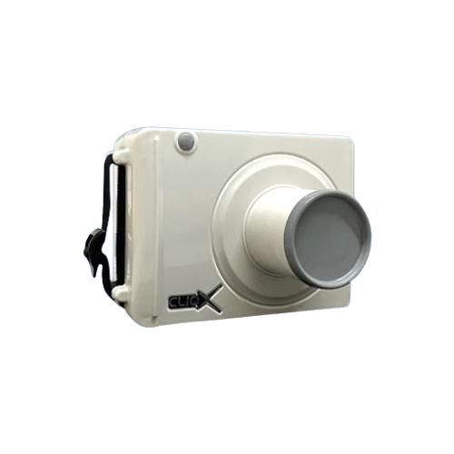
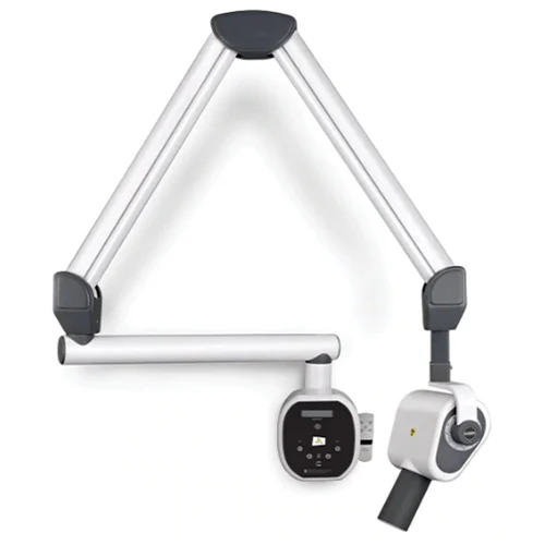
  1. We Are Engaged In Offering Dental X-Ray Machines Compatible With RVG (Radio Visio Graphy).

  2. This Product Is Also Known As MR-01 Wall Model Salient Features: Soft Touch Timing Selection.

  3. All Directional Movement With Angular Indication Enabling Perfect Positioning.

  4. Time Delay Of 10 Seconds Between Two Consecutive Exposures.

  5. Last Exposure Selection Retained In Memory. Lead Coated Radiography Long Cone.

  6. Exposure With Remote And Manual Also.

  7. Technical Specifications: X-Ray Output : 70kv,8ma Tube Filtration : 1.5mm Al Focal Point : 0.8mm Focal Skin Distance : 20cm Safety Leakage Radiation :<30mr/H At 1mtr. Distance Time Range : 0.01 To 3.0 Sec Line Voltage Compensation : AC 190v To 260v Powe Supply :230v, 50hz, 6A Exposure Time :Auto/Manual.


Packaging

1x X Ray Machine

1x Remote 

Product Data

  1. Power Supply

  2. 230V +- 10 %

  3. X-Ray tube

  4. 70kV, 10mA

  5. Focal spot

  6. 0.8mm x 0.8mm

  7. Focus on the skin distance 20 cm.

Filtration

  1. 1.5 –Al

  2. Exposure times- 0.1s to 3.0s

  3. Anatomic programs (Optional)

  4. 30 pre-set times with cordless remote

  5. Microprocessor-based timer with cordless remote for time setting and exposure.

Warranty
  1. 1 Year against Manufacturing Defect only

  2. Physical Damage is not covered in any case

  3. Service and Warranty are handled by the manufacturer/importer directly for this product.

Direction to Use

To Reduce Radiation Exposure, X-Ray Machines Aim The X-Rays At Only The Focus Area.

When X-Rays Come Into Contact With Our Body Tissues, They Produce An Image On A Metal Film.
Soft Tissue, Such As Skin And Organs, Cannot Absorb The High-Energy Rays, And The Beam Passes Through Them.

Parallel Technique

  1. The Film Is Placed Parallel To The Object Being Radiographed, And The Beam Positioned Perpendicular To Both The Sensor/Plate And The Tooth/Root.

  2. This Technique Results In The Most Accurate Image But Is Only Useful For The Caudal Mandibular Cheek Teeth Because:

  3. Dogs And Cats Do Not Have An Arched Palate And, Therefore, The Maxillary Teeth Cannot Be Imaged With This Technique

  4. The Mandibular Symphysis Interferes With The Placement Of The Sensor/Plate Parallel To The Tooth Roots Of The Mandibular Canines And Incisors As Well As The Rostral Mandibular Premolars.

  5. Bisecting Angle Technique

  6. This Technique Is Based On The Theory Of Equilateral Triangles And Creates An Image That Accurately Represents The Tooth And Roots.


Features
  1. The Sensor/Plate Is Placed As Parallel As Possible To The Tooth Roots.

  2. The Angle Between The Tooth Root And Sensor/Plate Is Measured Or Estimated.

  3. Then The Angle Is Cut In Half (Bisected) And The Beam Placed Perpendicular To This Bisecting Line.

  4. If The Angle Between The Sensor/Plate And The Beam Is Incorrect, The Radiographic Image Will Be Distorted Because The Beam Will Create An Image That Is Longer Or Shorter Than The Object Imaged.

  5. When The Angle Of The Beam To The Sensor/Plate Is:

Check Delivery Availability


Free shipping on all orders above ₹ 2000
10 Days Return Policy
View Policy
100% Genuine products

Suggest Us A Product


Have a product in mind that you'd like to suggest? Click the button below to submit your suggestion.


WhatsApp