Shop By Brands
 All Products
 Oralhealthcart Products
 Affordable Make in India Products

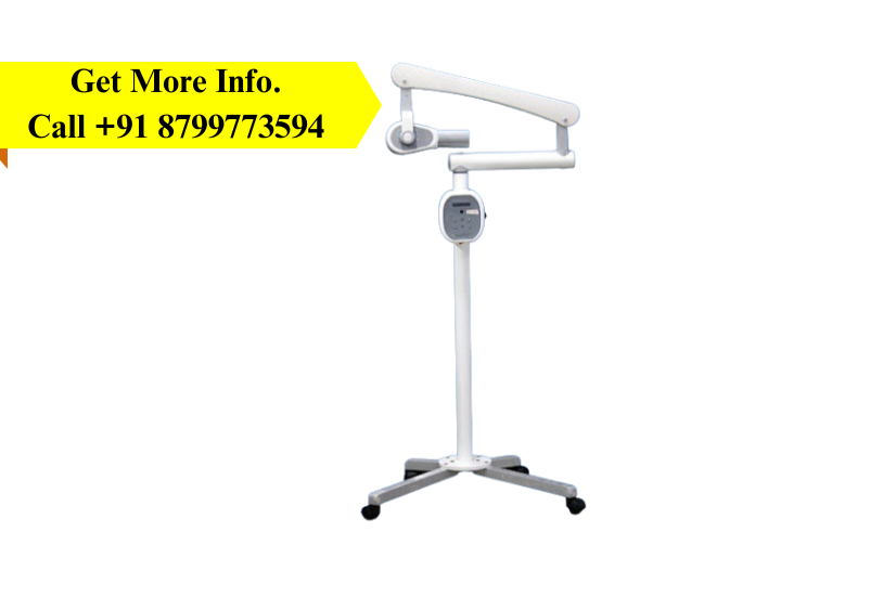
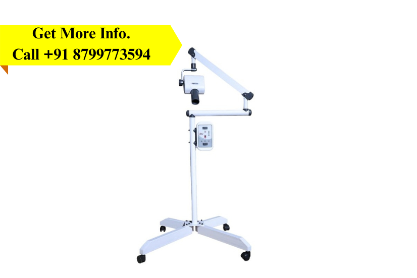
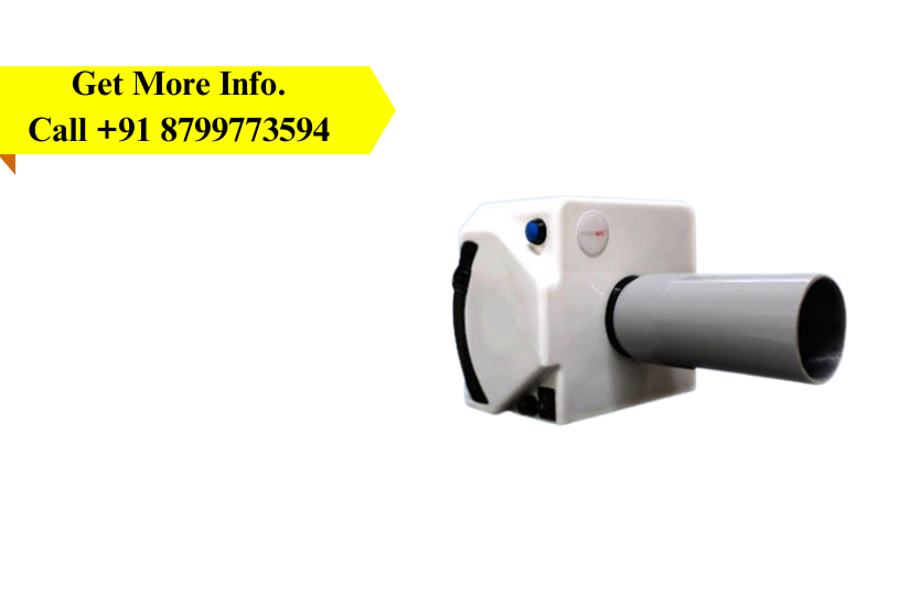
Description
-
We Are Engaged In Offering Dental X-Ray Machines Compatible With RVG (Radio Visio Graphy).
-
This Product Is Also Known As MR-01 Floor Model Salient Features: Soft Touch Timing Selection.
-
All Directional Movement With Angular Indication Enabling Perfect Positioning.
-
Time Delay Of 10 Seconds Between Two Consecutive Exposures.
-
Last Exposure Selection Retained In Memory.
-
Lead Coated Radiography Long Cone.
-
Exposure With Remote And Manual Also.